World's First Capsule Endoscope, PillCam sets world record
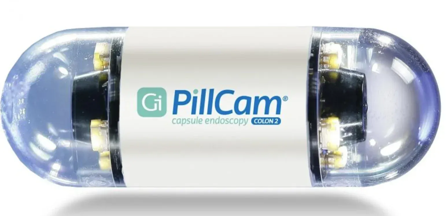
Minneapolis, Minnesota, United States--The mini video camera for the stomach, known as the PillCam, is the medical breakthrough pioneered the field of capsule endoscopy, setting the world record as the World's First Capsule Endoscope, according to the WORLD RECORD ACADEMY.
The Israeli mini video camera for the stomach is known as the PillCam. Invented in Israel by missile defense engineer Gavriel Iddan and gastroenterologist Eitan Scapa in the mid-1990s, the medical breakthrough pioneered the field of capsule endoscopy.
- How It Works
• The Device: The capsule is roughly the size of a large vitamin or jellybean (about 26 mm long and 11 mm in diameter). It contains a micro-video camera, LED lights, a radio transmitter, and a battery. - The Procedure: The patient swallows the capsule with water. It travels naturally through the digestive tract via regular muscle contractions (peristalsis), requiring no sedation and no invasive tubes.
- Data Collection: As it travels over an 8-hour period, it captures approximately 50,000 high-speed photos. It beams these images wirelessly to a recording device worn on a belt around the patient's waist.
- Disposal: The capsule is single-use and disposable; it is passed naturally out of the body and flushed away. (AI Overview)
Diagnostics and AI Integration
(AI Overview)
The primary purpose of the
PillCam is to map areas of the gastrointestinal tract that traditional upper endoscopies or colonoscopies cannot easily reach, specifically the 6-meter-long small intestine. Doctors use it to detect:
- Crohn's disease and celiac disease
- Gastrointestinal bleeding or unexplained anemia
- Polyps, tumors, and ulcers
Because reviewing 50,000 images is incredibly time-consuming, modern variations of this technology utilize artificial intelligence (AI) to quickly scan the photos and flag abnormalities for the physician.
Current Availability
The technology was commercialized by the Israeli company Given Imaging and received U.S. FDA approval in 2001. The company was later acquired, and the platform is now owned and distributed globally by
Medtronic under products like the
PillCam SB.
"Israel’s PillCam revolutionized GI imaging, and now others are following that success with other non invasive solutions that journey through the body. Every list of the greatest Israeli inventions includes PillCam,"the
Technion UK says.
"PillCam is a video camera swallowed like a vitamin pill. It travels through the gastrointestinal tract, sending clear images to the physician on its way out of the body. Invented by an Israeli electro-optical engineer and his neighbor, a gastroenterologist, the camera-in-a-pill endoscopy device signaled a worldwide diagnostic revolution. Capsule endoscopy has been FDA-approved and in use since 2001.
"The PillCam capsule endoscopy platform now enables physicians to detect GI abnormalities, monitor disease (such as Crohn’s) and assess treatment efficacy for conditions in the esophagus, stomach, small bowel and colon (large intestine)."
"Researchers at Given Imaging (Yoqneam, Israel) incorporated a digital camera, battery, radio transmitter, and light source into a pill-sized package that can be swallowed by patients to enable nonsurgical imaging of the gastrointestinal tract.
"The torpedo-shaped capsule can provide more than five hours of real-color images. The patient need not be in a hospital or clinic during this time. The disposable capsule, which is 30 mm long and 11 mm in diameter, is propelled by peristalsis through the gastrointestinal tract. The images are transmitted from the capsule using UHF signals to an antenna array attached to the patient (much like an ECG test). The aerials also record position information. The received images are stored in a recorder worn on a belt around the waist," the SPIE says.
"The data is later downloaded to a computer workstation and processed to produce a 20-minute video clip of the images transmitted by the capsule. The data also indicates where in the abdomen the capsule was at the time; this is important for locating diseased tissue should surgery be necessary."
"Capsule endoscopy is a medical procedure used to record internal images of the gastrointestinal tract for use in disease diagnosis. Newer developments are also able to take biopsies and release medication at specific locations of the entire gastrointestinal tract. Unlike the more widely used endoscope, capsule endoscopy provides the ability to see the middle portion of the small intestine. It can be applied to the detection of various gastrointestinal cancers, digestive diseases, ulcers, unexplained bleedings, and general abdominal pains. (Wikipedia)
"After a patient swallows the capsule, it passes along the gastrointestinal tract, taking a number of images per second which are transmitted wireless to an array of receivers connected to a portable recording device carried by the patient. General advantages of capsule endoscopy over standard endoscopy include the minimally invasive procedure setup, ability to visualize more of the gastrointestinal tract, and lower cost of the procedure.[2][medical citation needed]
History
"Capsule endoscopy was first conceptualized by Israeli engineer Gavriel Iddan and Israeli gastroenterologist Eitan Scapa in Boston, Massachusetts, in the early 1980s. The two partners initially developed a CCD (charge-coupled device) camera-based imaging system using a fiber-optic tether, but the prototype suffered from high power consumption and slow image transmission."
"